![New Vitamin A Finding Rewrites Vision Science [Revealed]](https://giglegravity.site/wp-content/uploads/2026/07/New-Vitamin-A-Finding-Rewrites-Vision-Science-Revealed.jpg)
Vitamin A and Thyroid Hormones Shape Central Vision Before Birth
Scientists at Johns Hopkins University have discovered how the human eye develops sharp central vision before birth. Their research shows that a vitamin A-derived molecule works with thyroid hormones at carefully controlled stages of retinal development.
This interaction helps the retina produce specialised light-sensing cells, known as photoreceptors, which allow people to see fine details, recognise faces and read clearly. The findings challenge a long-standing scientific explanation of how these important cells form.
Researchers made the discovery using retinal organoids, which are small, laboratory-grown tissues that closely imitate the developing human retina. The study may improve understanding of vision development and support future treatments for eye conditions such as macular degeneration, glaucoma and other diseases that damage the retina.
The research was published in the Proceedings of the National Academy of Sciences.
Scientists Reveal How the Retina Develops Its Sharpest Vision
Researchers at Johns Hopkins University have taken an important step towards understanding how the centre of the human retina develops. This specialised area is essential for clear central vision and is often the first part of the eye damaged by macular degeneration.
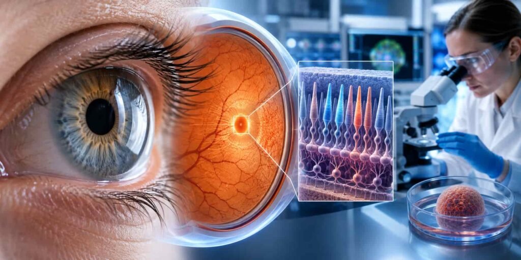
Robert J. Johnston Jr., an associate professor of biology who led the study, explained that a better understanding of this region could help scientists create more realistic retinal organoids. In the future, these laboratory-grown tissues may support the development of retinal transplantation methods designed to restore lost vision.
To study early eye development, the researchers used organoids, which are small clusters of tissue grown in the laboratory. These tissues closely copy important features of the developing human retina. By observing them for several months, the team identified the biological processes involved in forming the foveola.
The foveola is a tiny area at the centre of the retina that produces the sharpest and most detailed vision. Although it covers only a small part of the retina, it contributes to nearly half of human visual perception.
How Cone Photoreceptors Create Colour Vision
The research focused on cone photoreceptors, which are light-sensitive cells responsible for daylight vision, fine detail and colour vision. As the retina develops, these cells become blue, green or red cones. Each cone type responds to a different range of light wavelengths.
Most areas of the retina contain all three types of cones. However, the foveola contains mainly red cones and green cones, with no mature blue cones. Scientists have long been uncertain about how this unusual pattern develops.
Studying this process has been difficult because common laboratory animals, including mice and fish, do not form the same arrangement of cone cells as humans. The use of human retinal tissue models allowed the researchers to examine developmental changes that cannot be accurately studied in these animals.
Blue Cones Change During Fetal Development
The findings indicate that the specialised cone pattern of the foveola forms through a carefully controlled sequence during early fetal development.
Between weeks 10 and 12 of pregnancy, a small number of blue cone cells appear in the developing foveola. By approximately week 14, these cells have changed into red and green cones.
The researchers identified two biological mechanisms behind this transformation.
First, retinoic acid, a substance produced from vitamin A, is broken down in the developing retina. This reduces the production of additional blue cones.
Next, thyroid hormones signal the remaining blue cones to change into red and green cones. This coordinated process creates the cone arrangement needed for sharp central and colour vision.
The discovery provides new insight into photoreceptor development, retinal biology and the formation of the human foveola. It may also support future research into treatments for vision loss, macular degeneration and other diseases affecting the central retina.
Hormones Guide the Formation of Sharp Central Vision
The study shows that retinoic acid and thyroid hormones work in a carefully timed sequence during retinal development.
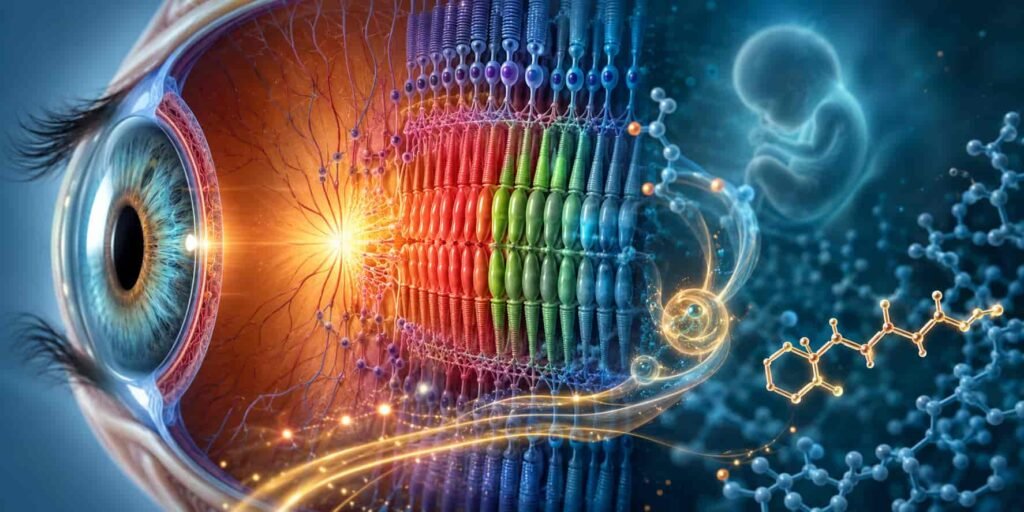
First, retinoic acid, which is produced from vitamin A, helps organise the early pattern of cone photoreceptors. Thyroid hormones then change the remaining blue cone cells into red and green cones.
According to Robert J. Johnston Jr., this transformation is important because blue cones in the central retina could reduce the quality of detailed vision. The correct balance of cone cells allows the foveola to support sharp eyesight and accurate colour vision.
New Evidence Changes an Earlier Scientific Explanation
For nearly 30 years, many researchers believed that blue cones formed in the centre of the retina and later moved towards its outer regions. Under that theory, each cone cell developed a fixed identity that remained unchanged.
The latest findings suggest a different process. Instead of moving away, the blue cones may stay in the same location and gradually change into red and green cones.
The researchers explained that more evidence is still needed to completely dismiss the earlier theory. However, their results strongly support cone cell conversion as an important part of foveal development.
This discovery provides a new explanation for how the human retina creates the specialised arrangement of photoreceptors needed for high-resolution vision.
Retinal Organoids Could Support Future Treatments
The research may also contribute to future treatments for vision loss. Johnston’s team is working to develop more advanced retinal organoids that closely reproduce the structure and function of the human retina.
These laboratory-grown tissues could help researchers create healthy and carefully selected populations of photoreceptor cells. In the future, these cells may be used in cell replacement therapy to repair damaged parts of the eye.
The aim would be to introduce healthy photoreceptors that could connect with existing retinal tissue and possibly restore some lost vision. This approach may be especially valuable for conditions such as macular degeneration, which damages central vision and currently has no complete cure.
Researchers stress that this technology remains at an early stage. Extensive testing will be required to confirm its safety, effectiveness and ability to work inside the human eye before clinical treatment becomes possible.
Even so, the findings provide a promising foundation for future research in regenerative medicine, retinal transplantation, photoreceptor replacement and vision restoration.
Read Also: AI Vaccine Clears First Human Test [Breakthrough]Summary: New Vitamin A Finding Rewrites Vision Science [Revealed]
Scientists at Johns Hopkins University found that retinoic acid, derived from vitamin A, and thyroid hormones guide the development of sharp central vision before birth.Using retinal organoids, they observed blue cone cells changing into red and green cones in the foveola.This challenges the older theory that blue cones simply moved away from the centre of the retina.The discovery improves understanding of photoreceptor development and colour vision.It may also support future treatments for macular degeneration, vision loss and retinal cell replacement.